Double Collecting System Kidney Ultrasound
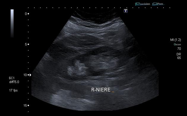
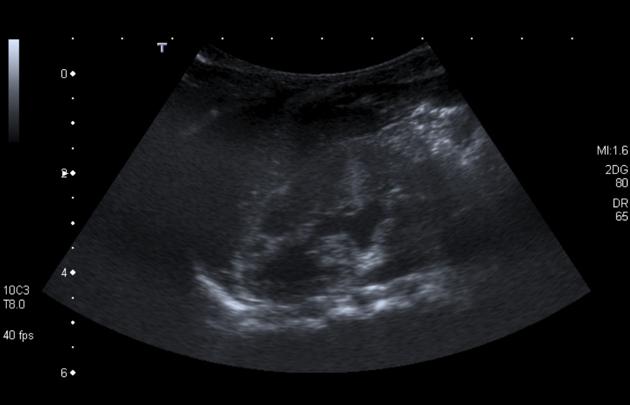
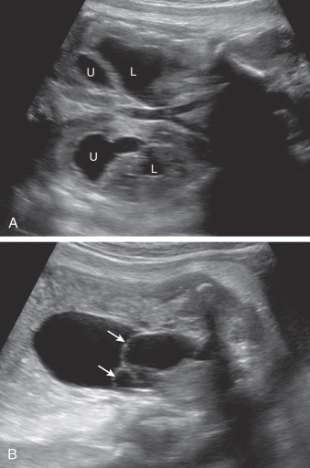
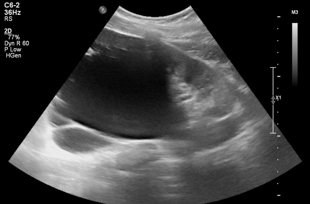
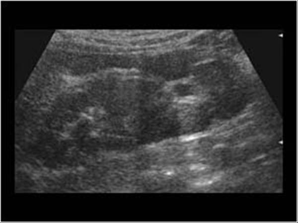
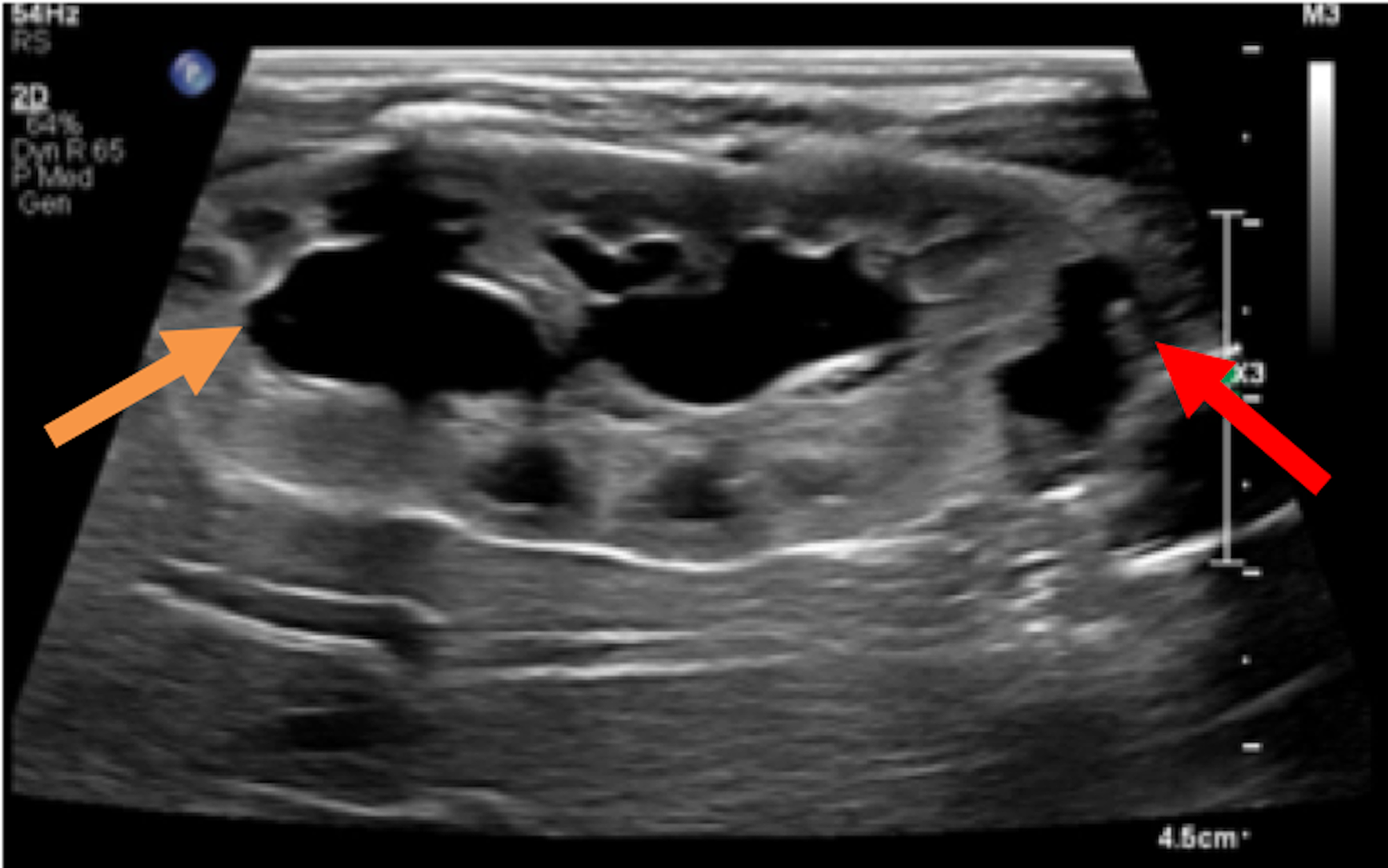
Double collecting system kidney ultrasound. It is characterized by an incomplete fusion of upper and lower pole moieties resulting in a variety of complete or incomplete duplications of the collecting system. Tubular stucture cranio-lateral to the bladder which is free of echos. Image shows an echo free structure on the right side retrovesically.
From about 9 to 12 weeks of pregnancy the fetal kidneys and adrenal glands can be visualized at both sites of the lumbar spine. - view Ultrasound 1 Ultrasound 1. Duplicated collecting systems also known as duplex collecting systems can be defined as renal units containing 2 pyelocaliceal systems that are associated with a.
Lateral kidney bulge same echogenicity as the cortex Hypertrophied column of Bertin. No evidence of obstructive hydronephrosis in either moiety. Indicated tortuosity no movement.
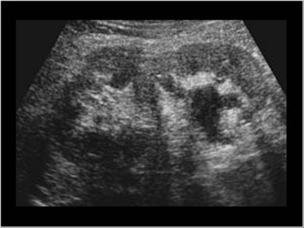
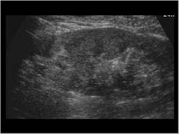
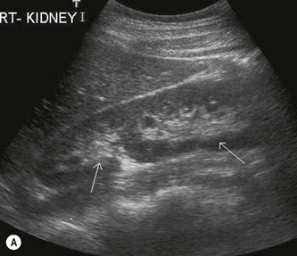
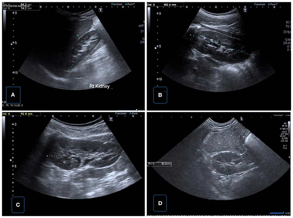
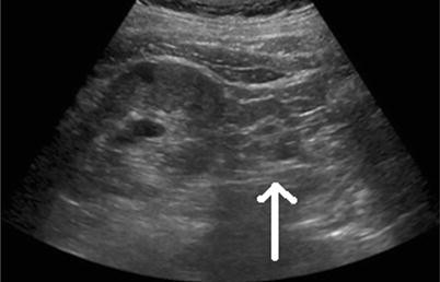
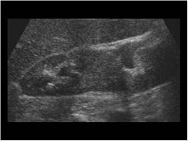
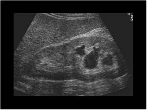
Greyscale ultrasound shows the typical appearance of a duplex kidney with two echo complexes and intervening cortical tissue on the right side. A duplex collecting system or duplicated collecting system is one of the most common congenital renal tract abnormalities. Duplication of the renal collecting system is recognized in approximately 1.
It disproportionately affects the left kidney and is bilateral in 15 to 20 of cases. The ectopic ureter and its orifice inserts medially and inferiorly to the ureter of the lower pole moiety and frequently ends as a ureterocele it can also insert into the urethra vagina etc. - view Ultrasound 2 Ultrasound 2.
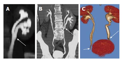
To investigate the clinical value of contrast-enhanced ultrasound CEUS in percutaneous nephrolithotomy PCNL for kidney stone patients without hydronephrosis. Thus this is a duplex right kidney with complete duplication of the collecting system double right ureters. The color Doppler ultrasound image shows a resultant double right ureteric jet emanating from duplication of the right ureteric openings.
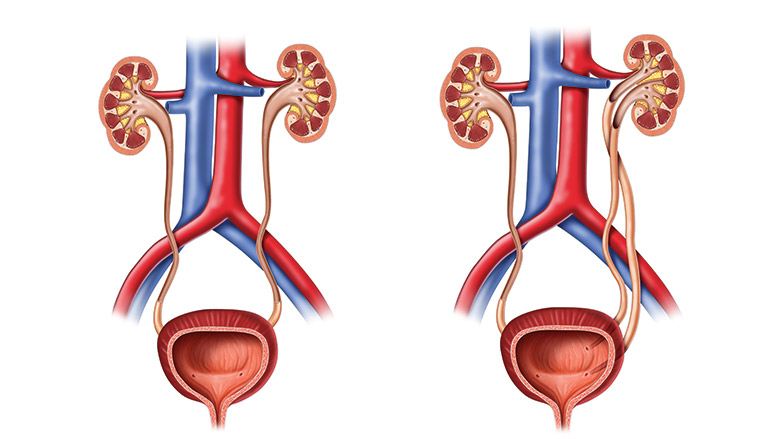
According to the rule the ureter of the upper pole moiety obstructs. Duplicated ureter or Duplex Collecting System is a congenital condition in which the ureteric bud the embryological origin of the ureter splits or arises twice resulting in two ureters draining a single kidneyIt is the most common renal abnormality occurring in approximately 1 of the population.
It disproportionately affects the left kidney and is bilateral in 15 to 20 of cases.
Greyscale ultrasound shows the typical appearance of a duplex kidney with two echo complexes and intervening cortical tissue on the right side. It disproportionately affects the left kidney and is bilateral in 15 to 20 of cases. Ultrasound US may show dilatation of one or both renal pelves with an intervening band of renal tissue. Children with this condition often also have a ureterocele an enlargement of the portion of the ureter closest to the bladder due to the ureter. Duplex kidneys can occur in one or both kidneys. Left kidney appeared normal. The objective of our study was to examine the risk for chromosomal aberrations in this isolated prenatal sonographic finding. - view Ultrasound 1 Ultrasound 1. From about 9 to 12 weeks of pregnancy the fetal kidneys and adrenal glands can be visualized at both sites of the lumbar spine.
Renal Ultrasound Basic Principles and BMUS Study Case Dromedary humps. Data from all chromosomal microarr. - view Ultrasound 3. Evaluation of the kidneys is part of the routine ultrasound examination done by many obstetricians as part of their prenatal care around the 20th week of pregnancy. Lateral kidney bulge same echogenicity as the cortex Hypertrophied column of Bertin. In rare instances duplex kidneys can appear in adults leading to diagnostic challenges. - view Ultrasound 2 Ultrasound 2.
















Post a Comment for "Double Collecting System Kidney Ultrasound"